53. Short muscles of foot
Anatomy
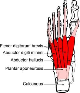
Flexor digitorum brevis (medial calcaneus & tubercle to middle phalanx of lateral four toes).
Flexor hallucis brevis (cuboid & third cuneiform to base of proximal phalanx & abductor tendon).
Extensor digitorum/hallucis brevis muscle (upper calcaneus on dorsum of foot distal to sinus tarsi to upper surface of toes).
Abductor hallucis (medial calcaneus to medial base of big toe).
Adductor hallucis (metatarsals & MT joints to lateral base of big toe). Important for the internal strength of the foot.
Interossei (metatarsals to base of proximal phalanx of toes)
Action - adducts toes, flexes proximal & extends distal toes 3-5.
Plantar aspect
The dome of calcaneus flares outward at it base. The medial tubercle is the weight bearing medial part. As well as serving as the attachment for muscle the medial tubercle serves as the attachment for the plantar aponeurosis. The plantar aponeurosis lies superficially and acts as a tie-beam to support the longitudinal arch of the foot. From the medial calcaneal tuberosity it fans out to attach on the metatarsal heads. The skin is thickened and supported with subcutaneous fibro-fatty tissue.
Heel spurs and plantar fasciitis can develop at or near the medial tubercle and will be tender. The treatment is to correct the biomechanics, which caused the problem and remove any further weight being placed on the spur with an orthotic. The orthotic must have a hole cut out of it for the spur to sit in. The patient should self treat by soaking their foot in a bath of warm water for five or ten minutes and then perform transverse friction over the plantar fascia for five minutes daily.
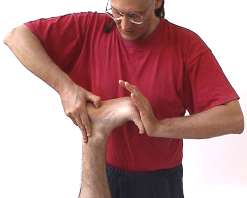
Technique 1 Kneading of short toe flexor muscles.
Patient
- Prone with their head rotated or straight and in a face hole.
Therapist
- Stand at the side of the table level with the patient's foot.
- Grasp the patient's ankle with your cephalad hand and flex their knee to 90 degrees.
- Grasp the patient's toes with your caudad hand and extend the patient's toes.
Applicator
- The tip of your thumb or a T bar.
 Picture 52.1
Tissue tension
- Take up the primary tissue tension with compression and a transverse force achieved by moving across the muscle with your thumb in a fixed flexed position.
- Your force is perpendicular to the muscle and in a medial-lateral direction.
- Longitudinal tension is taken up by extending the patient's toes.
Kneading
- Apply a rhythmical cycle of primary and longitudinal forces.
- Your pressure should be just short of discomfort.
- Start at the proximal attachment of the muscle and work down the sole of the foot.
- To relax the muscle work with the patients breathing cycle, about 15 breaths per minute.
- To stimulate increase your speed to about 40 cycles per minute.
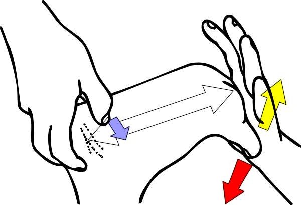 Figure 53.1
| Muscle Fibres | Primary Force | Secondary Force | Stretch Force | Contraction Force |
Active component
- Ask the patient to flex their toes and contract the short toe muscles for 3 to 5 seconds using a 10% effort.
- Resist the patient's effort and maintain tissue lockup throughout the technique.
- Ask the patient to exhale and to relax their muscle contraction.
- When you feel that the patient has relaxed their muscles completely (about 1-3 seconds) take up the tissue slack by increasing the primary and longitudinal tension.
- Take up the slack after each contraction and during exhalation.
- Repeat 2 or 3 times or as required.